

Animals
We used male C57BL/6 mice purchased from Jackson Laboratory Japan, Inc., and all mice were reared in a daylight cycle with a defined time from 8:00 AM to 8:00 PM in specific pathogen-free conditions. Male mice were used in this experiment because of the possibility that data may be scattered in female mice due to greater fluctuations in hormonal balance.
A COPD mouse model was developed by intratracheal PPE administration (E7885; Sigma-Aldrich, USA) followed by LPS (L3024; Sigma-Aldrich, U.S.A.)25. At 10 weeks of age (day 0), COPD mice models were intratracheally administered 0.375 U of PPE in 100 µL of PBS under isoflurane anesthesia. At 12 weeks of age (day 14), they were treated with 100 μg of LPS once daily for 3 days. Control mice were administered 100 μL of PBS instead of PPE and LPS (Fig. 8).
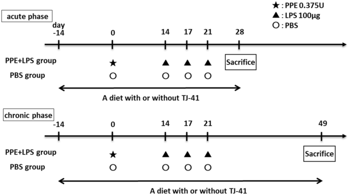
Study design for a COPD mice model. COPD mice model were administered PPE at 10 weeks of age (day 0), and they were treated with LPS three times every 3 days starting at 12 weeks of age (day 14). The 14-week-old mice (day 28) were considered an acute phase model of COPD exacerbation, whereas the 17-week-old mice (day 49) were considered chronic phase models of COPD.
A 2% TJ-41-containing or a control diet was fed to mice from 8 weeks of age until physiological and pathological analysis; 14-week-old mice (day 28) were considered the COPD acute exacerbation model, and 17-week-old mice (day 49) were considered the COPD chronic phase model (Fig. 8).
TJ-41
TJ-41 bulk powder was provided by Tsumura Co. (Tokyo, Japan). Further, 2% TJ-41 was added into rodents’ diets MF (Oriental Yeast Co., Ltd., Japan) for animal experiments, as previously described24,33. TJ-41 bulk powder was dissolved in ultrapure water for in vitro experiments. After adding Dulbecco’s Modified Eagle Medium (DMEM, WAKO, Japan) or RPMI-1640 medium (WAKO, Japan) at a ratio of 1:1, the solution was centrifuged, and the supernatant had a final concentration of TJ-41 of 10 μg/μL. The supernatant was added to the culture medium to achieve a final TJ-41 concentration of 50 μg/mL or 250 μg/mL because higher TJ-41 concentrations induced cell death in preliminary studies (data not shown).
Physiological analysis
After the intraperitoneal administration of combined triadic anesthesia to anesthetize the mice34, mice were intratracheally intubated with a blunted 18-gauge needle. It was then connected to SCIREQ flexiVent® (EMKA, Canada) to measure the physiological function of the mouse lung as a closed system. Airway resistance and lung compliance were measured three times, and each average value was adopted.
Bronchoalveolar lavage fluid analysis
After physiological analysis, 1 mL of sterile saline was injected through the needle using a syringe, the lungs were washed, and the lavage fluid was retrieved three times to obtain bronchoalveolar lavage fluid (BALF). The collected BALF was centrifuged at 450 × g for 10 min at 4℃. The pellet was dissolved in 1 mL of PBS after separating the supernatant, and cells were counted by a LUNA cell counter (Logos Biosystems, Korea). Diff-Quik staining (Sysmex, Japan) was used to measure leukocyte fractions of cells in BALF.
Lung histopathological analysis
Mouse lungs were fixed by intratracheal injection of 10% buffered formalin solution at a constant pressure of 25 cmH2O followed by immersion in formalin solution for at least 24 h. Specimens were embedded in paraffin, cut into 5-μm sections, and stained with hematoxylin–eosin.
Cell culture
We used two cell types for our in vitro experiments, BEAS-2B cells and U-937 cells, both provided by the Japanese Collection of Research Bioresources Cell Bank. BEAS-2B cells, which is a human epithelial cell line, were grown in DMEM (high glucose) with L-glutamine medium (WAKO, Japan) containing 10% fetal bovine serum (FBS). U-937 cells, which is a human macrophage cell line, were grown in RPMI-1640 with L-glutamine medium (WAKO, Japan) containing 10% FBS. Passages were limited to up to 20 times in each cell.
BEAS-2B cells were seeded in an FBS-free DMEM medium for starvation for in vitro experiments of LPS administration, and TJ-41 was administered at 50 μg/mL or 250 μg/mL. LPS was added at a final concentration of 5 µg/mL after incubation for 24 h. After 2 h, cells were retrieved for mRNA analysis. Starvation was performed in FBS-free RPMI-1640 with L-glutamine medium for U-937 cells, and thereafter, the same procedure was performed for BEAS-2B cells. In vitro studies of LPS administration have reported LPS concentrations of 100–10 μg/mL35,36. In particular, 5 μg/mL has been recognized as a highly effective concentration with minimal cell death37. Therefore, we decided to administer LPS at a concentration of 5 μg/mL in our study.
BEAS-2B or U-937 cells were starved and treated with TJ-41 solution at a final concentration of 250 μg/mL in time course analysis. The pAMPK, tAMPK, pS6K, and tS6K protein expressions were examined between 5 and 60 min after TJ-41 administration, and Nrf2 and LC3B protein expression was examined between 1 and 6 h after TJ-41 administration.
Quantitative real-time reverse transcription polymerase chain reaction (RT-PCR)
Cell pellets were lysed with TRIzol (Invitrogen, U.S.A.). Mouse lung tissue was also lysed in TRIzol followed by homogenization using an MS-100 bead homogenizer (Tomy, Japan). After extracting the total RNA, a micro-spectrophotometer was used to confirm that the A260/A280 absorbance of the isolated RNA was > 1.8. From 1.0 µg of total RNA, single-stranded cDNA was synthesized using ReverTraAce (Toyobo, Japan). Quantitative real-time RT-PCR was performed using Thermal Cycler Dice® Real-Time System III and TB Green Fast qPCR Mix (TaKaRa Bio, Japan) according to the manufacturer’s instructions. Quantification was performed in duplicate and the expression levels of target genes were calculated based on Gapdh mRNA levels using the delta-delta CT method. Supplementary Table 1 shows the primer sequences.
Western blotting analysis
Cell pellets were lysed in RIPA buffer (Wako, Japan) with protease inhibitor (Thermo Fisher Scientific, Japan) and phosphatase inhibitor (Thermo Fisher Scientific, Japan). Further, mouse lung tissue was lysed in this buffer followed by homogenization using an MS-100 bead homogenizer (Tomy, Japan). Thereafter, the lysate was centrifuged and the supernatant was collected. The supernatant protein concentration was measured by the BCA method (TaKaRa Bio, Japan), and the protein content of each sample was equalized to SDS (TCI, Japan). After SDS, proteins were electrophoresed, transferred to the membrane, and blocked with ECL prime blocking reagent (GE Healthcare, U.K.). The membrane, as primary antibodies, was incubated overnight in blocking reagent containing pAMPK (50,081; CST, U.S.A.), tAMPK (ab80039; abcam, U.K.), pS6K (9234; CST, U.S.A.), tS6K (2708; CST, U.S.A.), Nrf2 (16,396-I-AP; proteintech, U.S.A.), or LC3B (3868 s; CST, U.S.A.), respectively. The following day, the membrane was incubated for 1 h with the corresponding rabbit IgG antibody (ab6721; abcam, U.K.) or mouse IgG antibody (ab97023; abcam, U.K.), as the secondary antibody. Ez-Capture MG (ATTO, U.S.A.) was used to detect protein bands, and CS Analyzer 3.0 (ATTO, U.S.A.) was utilized to measure band absorbance.
RNA sequencing
Total RNA was extracted from mice treated with PBS and PPE and LPS using RNeasy Mini Kit (Qiagen, Netherlands) following the manufacturer’s instructions. CLC Genomics Workbench software (Qiagen, Netherlands) was used for RNA sequencing (RNA-seq) analysis. R software was used for statistical calculations, and edgeR software38 was used for differential expression analysis. The DEG thresholds were defined as log2 fold change of > 1 or < 1 and a false discovery rate of < 0.05. Gene ontology analysis was performed using the Enrichr combined score39. The data set was stored in the Gene Expression Omnibus database (GSE239537).
Study approval
All mouse experiments were approved by the Ethics Committee for Animal Experiments of The University of Tokyo (P17-006), and were performed in accordance with ARRIVE guidelines and regulations of this committee. In other experiments, all methods were performed according to relevant guidelines and regulations. This study used no human specimens.
Statistics
Data was presented as means ± standard errors of the mean. GraphPad Prism 5 software (GraphPad Software Inc., USA) was used for statistical analyses. Unpaired t-tests were used for the analysis of two groups, and one-way analysis of variance and Tukey’s multiple comparison tests were used for the analysis of three or more groups unless otherwise stated. P-values of < 0.05 were considered statistically significant.